
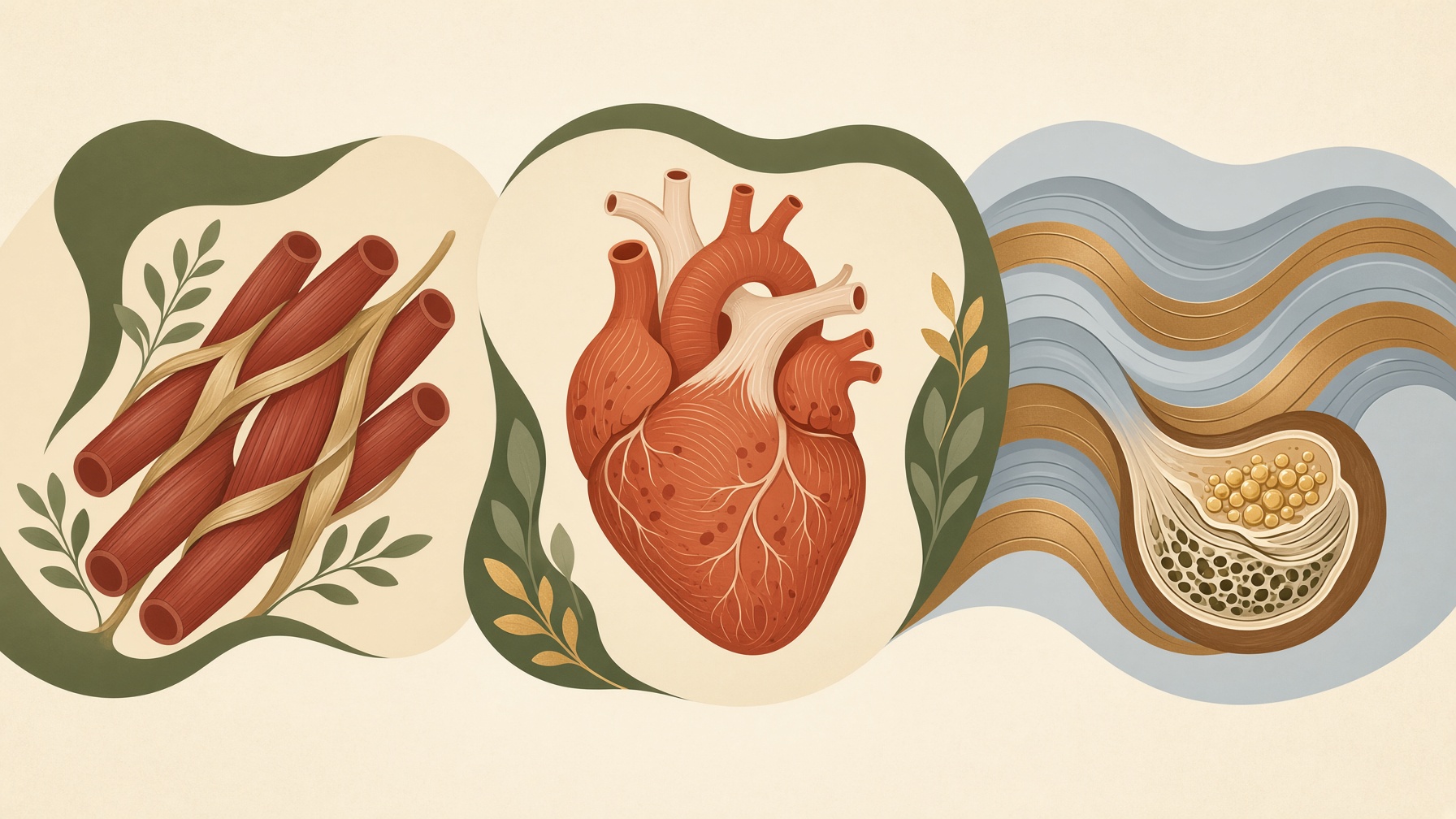
What Makes Skeletal, Cardiac, and Smooth Muscle Different?
Skeletal, cardiac, and smooth muscle share the ability to contract, but differences in cell structure and control suit each to different tasks.
Wikis
Intercalated discs are specialized regions of cell-to-cell contact that connect neighboring cardiac muscle cells, or cardiomyocytes. They are located mainly where the ends of adjacent cardiomyocytes meet and contain several types of junctions organized together. These connections allow heart muscle cells to remain mechanically linked during repeated contractions while also supporting rapid electrical communication between cells .1,2
Three major junctional components are traditionally recognized within intercalated discs: adherens junctions, desmosomes, and gap junctions. Adherens junctions connect the contractile apparatus of neighboring cells through the actin cytoskeleton, helping transmit mechanical force from one cardiomyocyte to the next. Desmosomes anchor intermediate filaments and provide strong cell-to-cell adhesion that helps the tissue withstand mechanical stress .1,2
Gap junctions provide a different type of connection. They contain channels formed by proteins called connexins that connect the cytoplasm of neighboring cardiomyocytes. Ions and other small molecules can pass through these channels, providing electrical coupling that helps electrical activity propagate from cell to cell and coordinates contraction across the myocardium .1,2
An intercalated disc is therefore not a single type of junction. It is an organized junctional region in which mechanical and electrical connections interact closely. This integration helps cardiac muscle function as a mechanically connected and electrically coordinated tissue .1,2
From this collection
Skeletal, cardiac, and smooth muscle share the ability to contract, but differences in cell structure and control suit each to different tasks.