
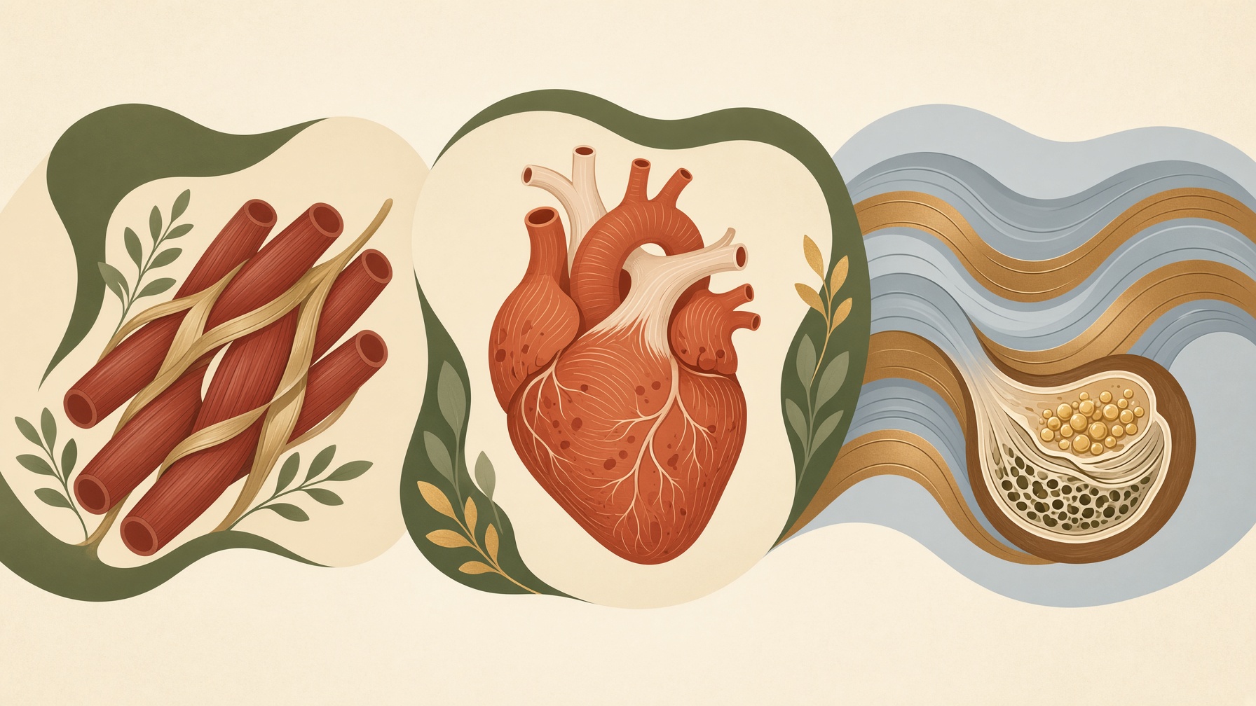
What Makes Skeletal, Cardiac, and Smooth Muscle Different?
Skeletal, cardiac, and smooth muscle share the ability to contract, but differences in cell structure and control suit each to different tasks.
Wikis
Smooth muscle is one of the three major types of muscle tissue, alongside skeletal and cardiac muscle. It is typically made of elongated, spindle-shaped cells that contain the contractile proteins actin and myosin. Unlike skeletal and cardiac muscle, smooth muscle does not have the repeating sarcomere organization that produces visible striations under a microscope, giving the tissue its “smooth” appearance .1
Smooth muscle is found mainly in the walls of hollow organs and tubes, including blood vessels, the digestive tract, urinary tract, reproductive tract, and airways. Its contractions can propel material through organs or alter the diameter and shape of a structure. For example, vascular smooth muscle changes blood-vessel diameter, while gastrointestinal smooth muscle contributes to movement of contents through the digestive tract .1
Smooth muscle contraction is generally not under conscious voluntary control. Its activity can instead be influenced by the autonomic nervous system, hormones, local chemical signals, changes in membrane electrical activity, and mechanical stretch. When intracellular calcium rises, calcium binds to calmodulin, which activates myosin light-chain kinase. This enzyme phosphorylates myosin light chains, promoting interactions between myosin and actin that generate force .1,2
This regulatory mechanism differs from contraction in skeletal muscle, where calcium acts primarily through the troponin system. Smooth muscle can also maintain force for relatively long periods with comparatively low energy use, a property suited to functions such as maintaining blood-vessel tone .1,2
From this collection
Skeletal, cardiac, and smooth muscle share the ability to contract, but differences in cell structure and control suit each to different tasks.