
What Makes Air Move In and Out of Your Lungs?
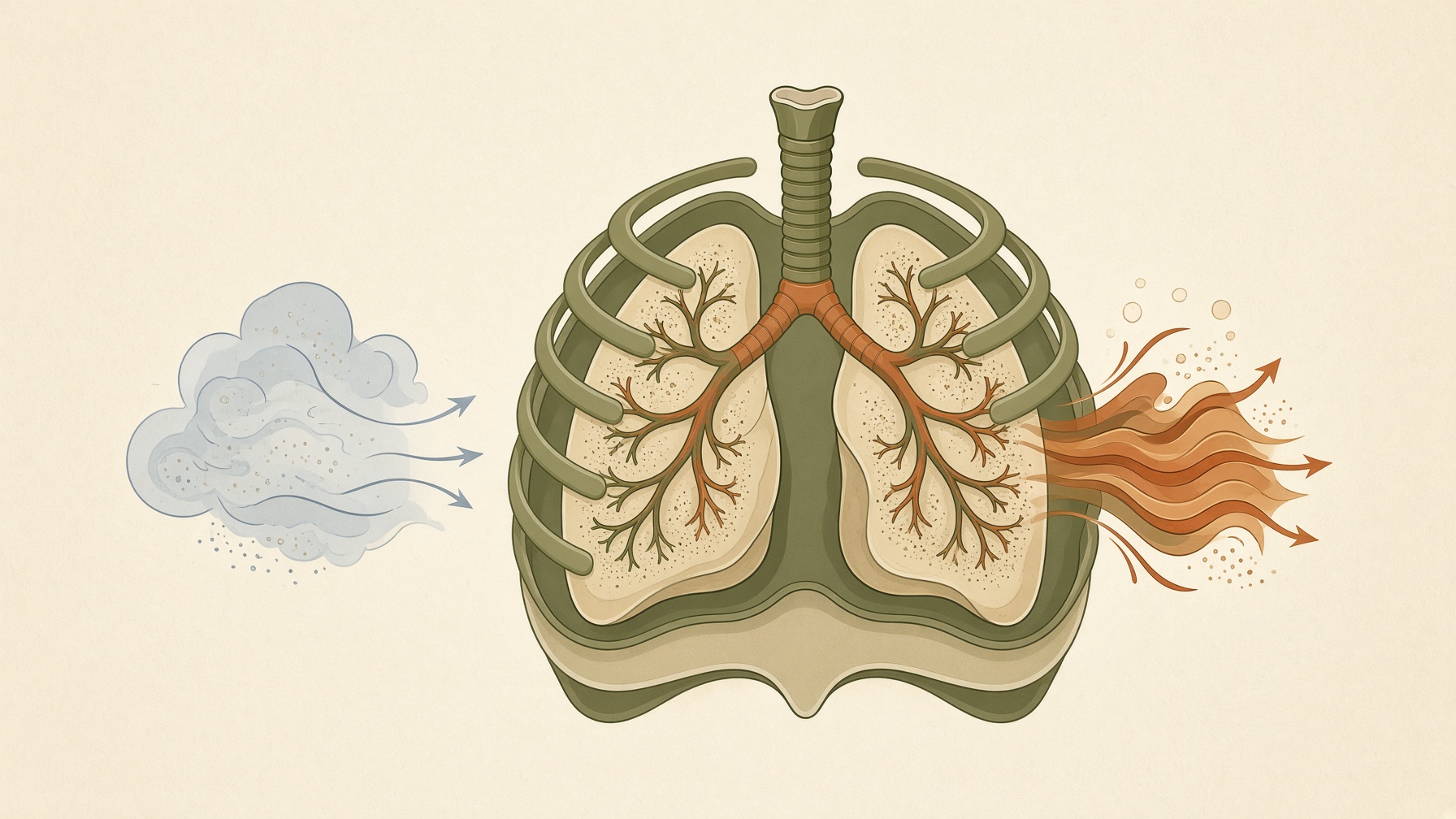
Breathing moves air through pressure differences created as the lungs expand and recoil, with airflow traveling from higher to lower pressure.
Wikis
The diaphragm is a dome-shaped sheet of skeletal muscle and tendon that separates the thoracic cavity, containing the lungs and heart, from the abdominal cavity. It forms the floor of the thorax and the roof of the abdomen and is the principal muscle of inspiration during normal breathing .1,2
The muscle fibers of the diaphragm arise around the lower opening of the thorax from the sternum, lower ribs and costal cartilages, and lumbar vertebral region. These fibers converge toward a broad central tendon. The diaphragm forms right and left domes, with the right dome generally positioned higher because of the liver beneath it .1
During inspiration, the diaphragm contracts and descends. This increases the vertical dimension of the thoracic cavity and makes intrapleural pressure more negative, contributing to expansion of the lungs and a fall in alveolar pressure that draws air inward. During quiet expiration, the diaphragm relaxes and rises as the respiratory system recoils toward its resting position .1,2
The diaphragm is not a completely solid barrier between the chest and abdomen. Several structures pass through or behind it. Its major passages accommodate the inferior vena cava, esophagus, and aorta. Motor control of the diaphragm is supplied primarily by the right and left phrenic nerves, which arise from cervical spinal nerve roots C3 to C5 .1
From this collection
Breathing moves air through pressure differences created as the lungs expand and recoil, with airflow traveling from higher to lower pressure.