
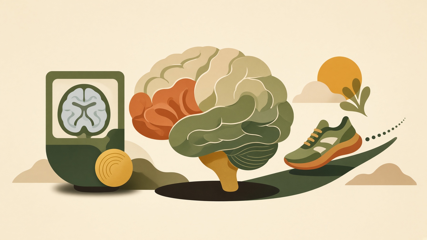
The Brain-Age Score Got Younger on the MRI. Did the Brain, or Just the Reading?
Exercise can shift some MRI-based brain-age estimates, but changes depend on the model and do not demonstrate literal reversal of brain aging.
Wikis
Functional MRI detects blood-related changes associated with neural activity, providing an indirect signal that also depends on vascular responses.
Functional magnetic resonance imaging, or fMRI, is an MRI-based method used to map changes in brain activity indirectly. The most common form uses the blood-oxygen-level-dependent, or BOLD, signal, which is sensitive to changes in the magnetic properties of blood as the balance between oxygenated and deoxygenated hemoglobin changes in active tissue .1
Neural activity increases local energy demand and triggers changes in blood flow, blood volume, and oxygen use. BOLD fMRI detects the resulting hemodynamic response rather than electrical activity from neurons themselves. Researchers can compare the signal during different tasks or examine spontaneous fluctuations at rest to study functional organization and connectivity. The signal is spatially detailed but develops more slowly than the underlying neural events .1,2
fMRI should therefore be distinguished from structural MRI. Structural MRI primarily depicts anatomy, such as gray matter, white matter, and lesions, while fMRI is designed to measure time-varying physiological signals related to function. A brighter or stronger BOLD signal is not a direct measure of how many neurons fired, and interpretation depends on vascular as well as neural factors. This indirect relationship is central to understanding both the power and limitations of fMRI .1,2
From this collection
Exercise can shift some MRI-based brain-age estimates, but changes depend on the model and do not demonstrate literal reversal of brain aging.