
How Are the Tiny Filaments Inside Your Muscles Arranged?
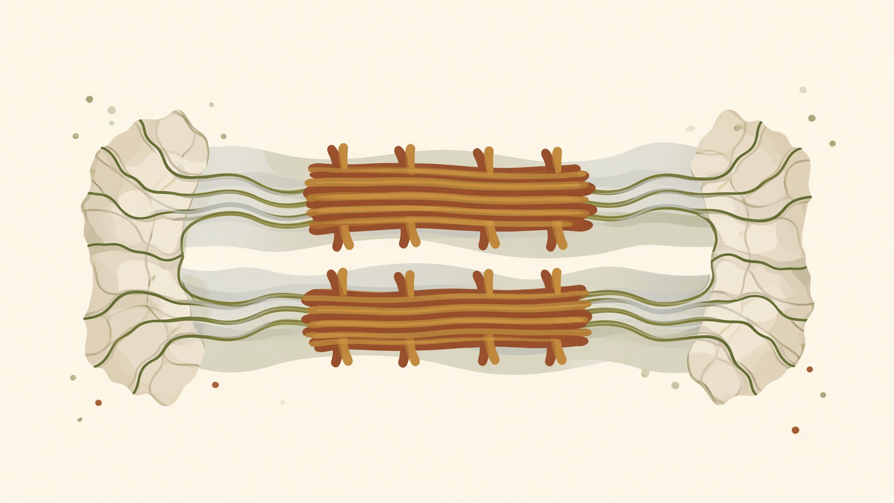
Sarcomeres shorten as thin filaments slide alongside thick filaments, increasing overlap without reducing the length of the filaments themselves.
Wikis
The M-line is a protein-rich structure at the center of a sarcomere, the repeating contractile unit of striated skeletal and cardiac muscle. It lies in the middle of the A band, within the central region occupied by thick, myosin-containing filaments. Its main structural role is to link and organize neighboring thick filaments, helping maintain their orderly arrangement within the sarcomere .1,2
Proteins of the myomesin family are major structural components of this region. Myomesin molecules interact with myosin and with titin, the giant protein that extends from the Z-disc toward the center of the sarcomere. Through these connections, the M-line is integrated with both the thick-filament lattice and the structural framework of the sarcomere .2
The terms M-line and M-band are sometimes used broadly for the same central sarcomeric region, although they can describe different levels of structure. Electron microscopy shows that the wider M-band can contain several narrow, electron-dense M-lines, including a central M1 line and additional lines on either side. Their prominence and protein composition can vary among muscle types .2
The M-line is distinct from the Z-disc. Both help organize sarcomeric filaments, but the M-line is located at the sarcomere center and primarily organizes thick filaments, whereas Z-discs form the sarcomere boundaries and anchor thin, actin-containing filaments .1,2
From this collection
Sarcomeres shorten as thin filaments slide alongside thick filaments, increasing overlap without reducing the length of the filaments themselves.