
Stretching may look like a simple change in muscle length, but the nervous system is receiving information throughout the movement. That sensory input helps the body keep track of what is happening mechanically and can influence the response that follows. Understanding this feedback also helps explain why stretching cannot be reduced to a simple rule that a muscle either contracts or relaxes when its length changes.
Muscle spindles and Golgi tendon organs are principal proprioceptive receptors associated with the muscle–tendon system. Their sensory roles are distinct. Muscle spindles provide information about muscle length and changes in length during stretch. Golgi tendon organs, by contrast, provide feedback primarily related to muscle–tendon force or tension. These two forms of information help distinguish what each receptor contributes to proprioceptive signaling .1
Muscle-spindle signaling can participate in the stretch reflex. When a muscle is stretched sufficiently, that stretch can evoke a reflex response that activates the stretched muscle. The size of that reflex depends on the characteristics of the stretch and the state of the nervous system, so a slow static stretch does not necessarily produce an obvious reflex contraction .1,2
Spindle-related signaling can also influence muscles that oppose the action of the stretched muscle. Activity from muscle-spindle Ia afferents can inhibit antagonist motoneurons through spinal reciprocal inhibitory pathways. This process is called reciprocal inhibition. The strength of this inhibitory effect can vary with the task and neural conditions, so reciprocal inhibition should not be interpreted as guaranteed relaxation of the antagonist during every stretch .2
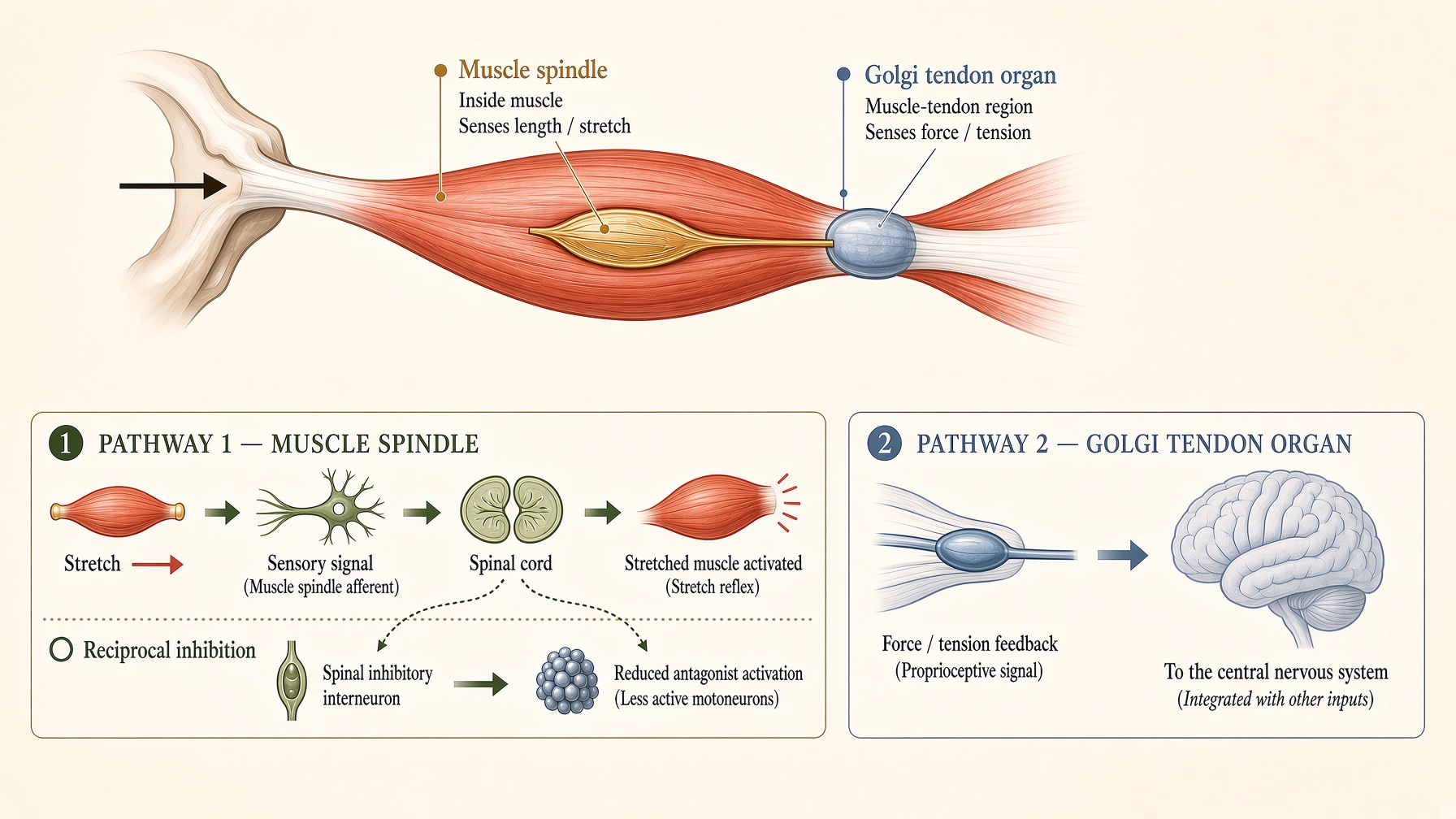
The practical value of separating these receptors is that it prevents stretching from being explained with overly simple reflex rules. Sensory information from muscles and tendons is combined with other spinal and supraspinal influences before a motor response emerges. Neither the muscle spindle nor the Golgi tendon organ acts as a simple on–off switch for contraction or relaxation. Their signals are part of a larger control system that continuously adjusts movement and muscle activity to the situation.

Join the discussion
No account or email is required. Your display name and comment will be public.