
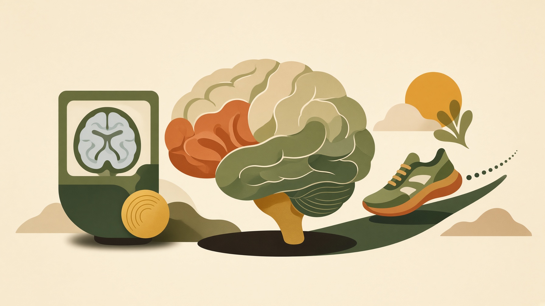
The Brain-Age Score Got Younger on the MRI. Did the Brain, or Just the Reading?
Exercise can shift some MRI-based brain-age estimates, but changes depend on the model and do not demonstrate literal reversal of brain aging.
Wikis
MRI creates tissue images from magnetic and radiofrequency signals without ionizing radiation, while still requiring specific safety screening.
Magnetic resonance imaging, or MRI, is a medical imaging technique that uses a strong magnetic field, radiofrequency energy, and magnetic-field gradients to generate detailed images of internal tissues. Most clinical MRI signals come from hydrogen nuclei, especially those in water and fat. The scanner detects signals emitted as excited nuclei return toward their equilibrium state .1
Image contrast can be changed by altering pulse sequences and timing so that differences in tissue properties, including T1 and T2 relaxation, contribute differently to the final image. Magnetic-field gradients encode the spatial origin of the signals, allowing a computer to reconstruct slices or three-dimensional volumes. MRI can therefore produce many types of soft-tissue contrast without using ionizing radiation .1,2
MRI is not the same as functional MRI, which uses particular MRI methods to infer changes related to brain activity. MRI also should not be described as risk-free simply because it lacks ionizing radiation. The strong magnetic and radiofrequency fields create specific safety considerations for metallic objects, some implants, heating, acoustic noise, and contrast agents when used. Screening and protocol selection are therefore integral parts of MRI practice .2
From this collection
Exercise can shift some MRI-based brain-age estimates, but changes depend on the model and do not demonstrate literal reversal of brain aging.
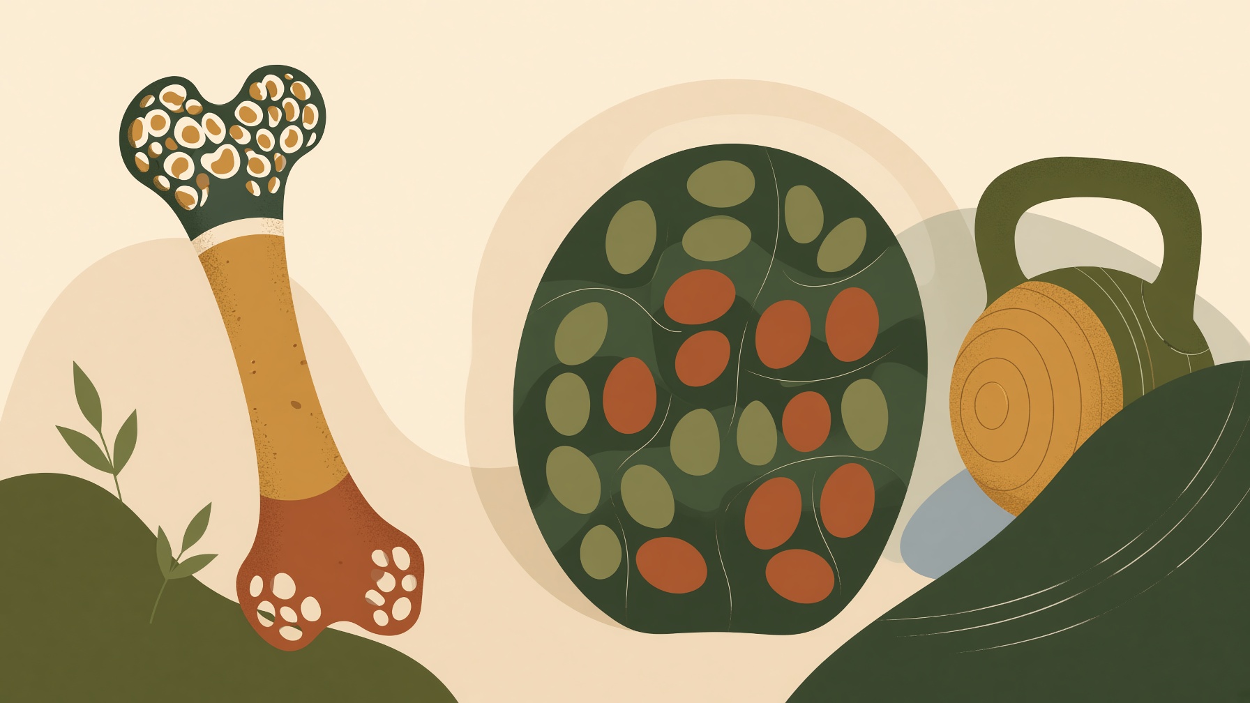
Exercise can improve bone density and muscle fat infiltration in studied groups, but these imaging measures describe different aspects of tissue health.
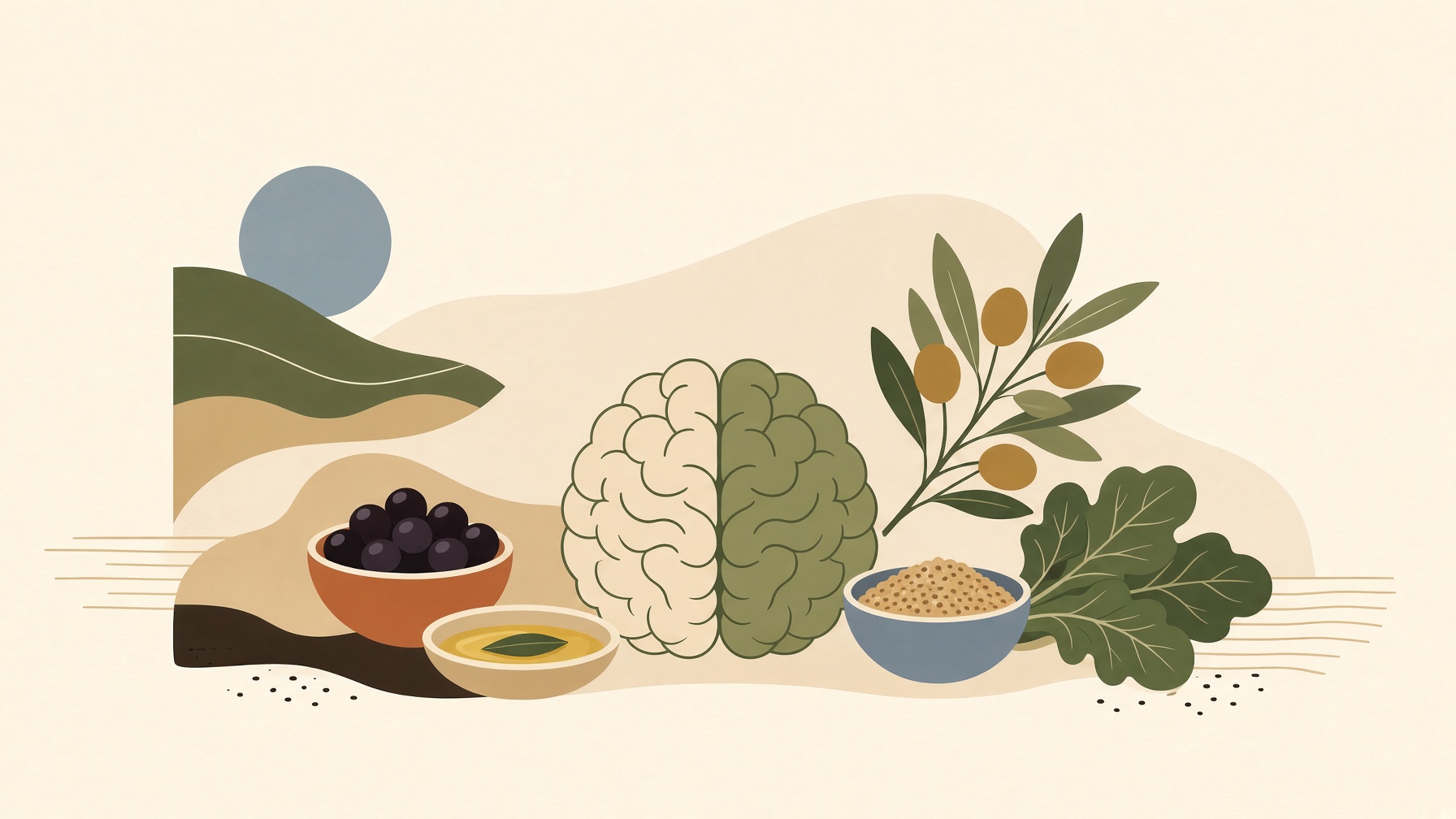
Brain-healthy dietary patterns show promising observational links, but trials do not establish one superior diet or a single food that protects cognition.