
How Are the Tiny Filaments Inside Your Muscles Arranged?
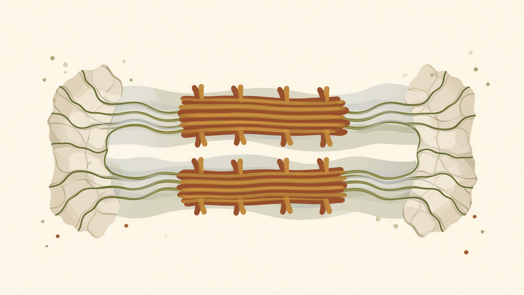
Sarcomeres shorten as thin filaments slide alongside thick filaments, increasing overlap without reducing the length of the filaments themselves.
Wikis
A sarcomere is the repeating contractile unit of striated muscle and the basic structural unit of a myofibril. Sarcomeres occur in skeletal and cardiac muscle and are arranged end to end along the length of each myofibril. A single sarcomere extends from one Z disc to the next, and the highly ordered arrangement of its proteins produces the characteristic striped appearance of striated muscle .1,2
The main contractile structures within a sarcomere are thin filaments, composed primarily of actin, and thick filaments, composed primarily of myosin. Thin filaments extend inward from the Z discs, while thick filaments occupy the central region and are organized around the M line. Their arrangement creates recognizable regions called the A band, I band, and H zone. Regulatory proteins including troponin and tropomyosin are associated with the thin filaments .1,2
During contraction, calcium binding to troponin helps permit interactions between myosin and actin. Myosin heads then undergo ATP-dependent cross-bridge cycling, generating force. When mechanical conditions allow the sarcomere to shorten, the thin filaments slide toward the center of the sarcomere. The filaments themselves do not substantially shorten; instead, their overlap increases and the Z discs move closer together .1,2
As a sarcomere shortens, the I band and H zone become narrower, while the A band remains essentially constant in length because the thick filaments retain their length. Coordinated shortening of many sarcomeres can contribute to shortening of the muscle fiber, while cross-bridge cycling can generate force whether or not substantial shortening occurs .1,2
From this collection
Sarcomeres shorten as thin filaments slide alongside thick filaments, increasing overlap without reducing the length of the filaments themselves.