
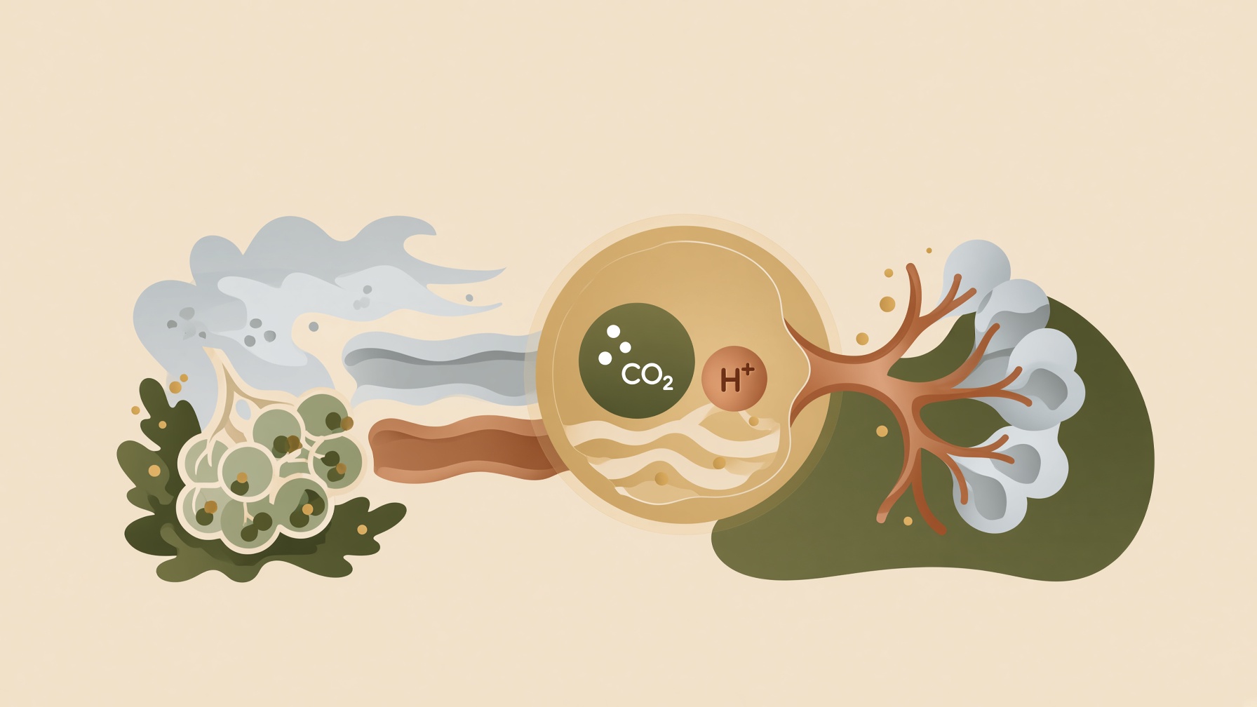
You rarely have to think about taking your next breath. As carbon dioxide, acidity, and oxygen levels change, the body can automatically adjust how deeply or how often you breathe. You can still take voluntary control for activities like singing or swimming underwater, but most breathing regulation happens without conscious attention.
Breathing usually occurs automatically, but it can also be consciously modified for voluntary behaviors. Automatic breathing is generated and regulated primarily by interacting respiratory networks in the brainstem, with additional influence from higher brain regions and sensory feedback. These networks send motor commands to the muscles that produce ventilation. The result is a system that can keep breathing going without conscious attention while still allowing voluntary changes when needed .1
Chemical feedback is an important part of that automatic regulation. Under normal oxygen conditions, carbon dioxide and hydrogen ions, or CO2/H+, are major chemical regulators of ventilation, although they are not the only influences on breathing. As arterial CO2 rises, carbon dioxide readily crosses the blood-brain barrier into brain extracellular fluid. The increase in CO2 shifts acid-base equilibria toward a higher hydrogen-ion concentration and lower pH. Increased brain CO2/H+ then stimulates central respiratory chemoreception and increases respiratory drive .2
As respiratory drive increases, ventilation can rise through a greater tidal volume, a higher breathing frequency, or both. The resulting increase in alveolar ventilation removes more carbon dioxide through the lungs, helping bring arterial CO₂ back toward its regulated range .2
Oxygen also affects ventilation, but its influence has a different pattern. Falling arterial PO₂ provides an increasingly strong stimulus to breathe as hypoxemia becomes more severe. Peripheral chemoreceptors, particularly the carotid bodies, are the principal sensors driving this response to reduced arterial oxygen. Their activity should not be thought of as switching on only at one exact value. Rather, the hypoxic ventilatory response becomes markedly stronger as arterial PO2 falls to around 60 mm Hg and below. That figure is an approximate inflection range, not a sharp physiological threshold .3
Most of this regulation happens without ever reaching your conscious attention. Rising carbon dioxide and acidity can push ventilation upward, while falling oxygen becomes a much stronger signal when arterial oxygen drops substantially. The result is a breathing system that continuously adjusts to changes inside the body, even when you are not thinking about breathing at all.
Join the discussion
No account or email is required. Your display name and comment will be public.