
How Your Body Digests and Absorbs Dietary Fat
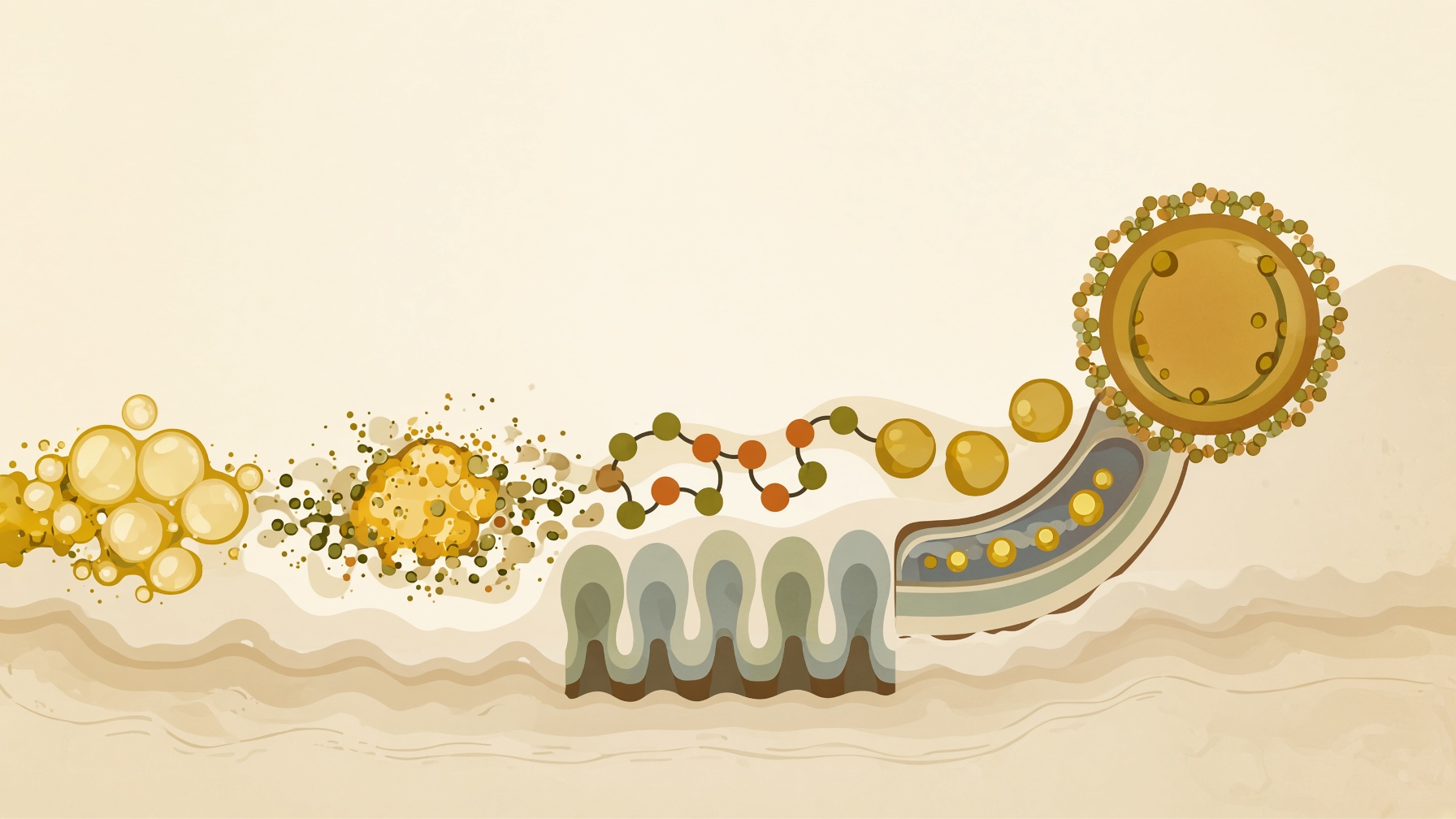
Dietary fat digestion combines physical dispersal by bile salts with enzyme breakdown, producing lipid components that intestinal cells can absorb.
Wikis
Enterocytes are specialized absorptive epithelial cells that line the intestinal surface, particularly the villi of the small intestine. They make up the majority of cells in the small-intestinal epithelium and form a major interface between material in the intestinal lumen and the body's internal environment. Enterocytes are polarized cells, meaning that their surface facing the intestinal lumen differs structurally and functionally from the surface facing underlying tissues and the circulation .1,2
The apical surface of an enterocyte is covered by densely packed projections called microvilli, which collectively form the brush border. Microvilli greatly increase the membrane area available for digestion and absorption. The brush-border membrane contains digestive enzymes and transport proteins involved in processing and taking up nutrients. Transporters on the basolateral membrane allow absorbed substances to leave the enterocyte and move toward underlying tissues and ultimately the blood or lymphatic circulation .1
Enterocytes participate in the absorption and processing of carbohydrates, amino acids and small peptides, lipids, vitamins, minerals, electrolytes, and water. Their functions vary along different regions of the intestine and along the crypt-to-villus axis. Enterocytes also contribute to the intestinal barrier by forming a continuous epithelial layer connected by intercellular junctions that regulate movement between neighboring cells .1,2
Enterocytes arise from intestinal stem cells located in crypts and differentiate as they move toward the intestinal surface. They are distinct from other intestinal epithelial cells such as mucus-producing goblet cells, hormone-secreting enteroendocrine cells, and Paneth cells, which have specialized secretory and defensive functions .2
From this collection
Dietary fat digestion combines physical dispersal by bile salts with enzyme breakdown, producing lipid components that intestinal cells can absorb.
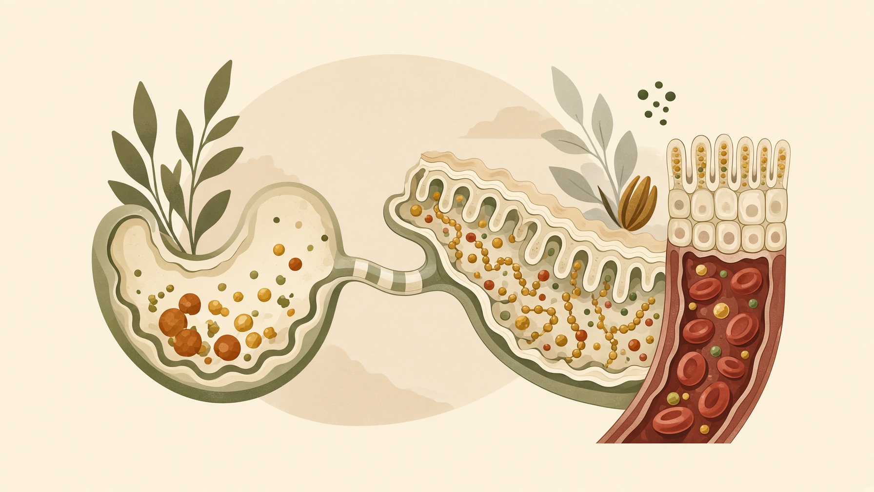
Protein digestion breaks food proteins into amino acids and small peptides that intestinal cells can absorb for the body’s ongoing protein turnover.