
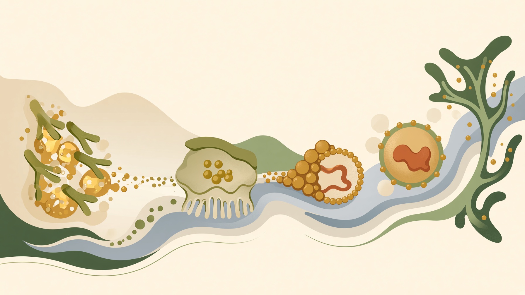
Dietary fat creates an unusual transport problem. Lipids are largely hydrophobic, yet digestion and circulation take place in environments dominated by water. The body cannot solve that problem by simply dissolving fat, so it relies on specialized structures and processes that allow lipids to interact with watery surroundings without behaving as freely dissolved molecules.
Most digestive enzymes operate in aqueous gastrointestinal fluids, whereas dietary fats are poorly water-soluble. In the small intestine, bile components, particularly bile salts and phospholipids, promote emulsification. This disperses dietary fat and increases the lipid-water interface, making lipid substrates more accessible to digestive enzymes. Pancreatic lipase then hydrolyzes dietary triglycerides mainly into free fatty acids and 2-monoglycerides. The result is a transition from relatively inaccessible dietary fat to smaller lipid products that can move into the next stage of intestinal processing .1,2
Those products still need help moving through the intestinal environment to the absorptive surface. Mixed micelles keep poorly water-soluble lipid digestion products dispersed in the intestinal contents and facilitate their movement through the aqueous environment near the brush border. At the absorptive surface, lipid molecules leave the micellar phase and are taken up by enterocytes; the bile salts themselves largely remain in the intestinal lumen for later reabsorption .1,3
Absorption does not simply send those lipid components onward in the same form. Inside enterocytes, absorbed long-chain fatty acids and monoglycerides are largely re-esterified to form triglycerides. These triglycerides are then packaged with other lipids and apolipoproteins into particles called chylomicrons. This packaging creates a form suited to moving substantial amounts of absorbed dietary lipid out of the intestine and toward the circulation .1
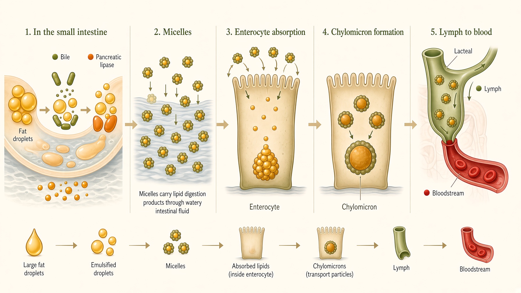
This pathway applies mainly to long-chain dietary fatty acids. Shorter-chain fatty acids, particularly those derived from medium-chain triglycerides, are handled differently and can pass into the portal circulation without first being packaged into chylomicrons .1
Newly formed chylomicrons are secreted from enterocytes and enter intestinal lymphatic capillaries called lacteals. They travel through the lymphatic system and ultimately enter the bloodstream, largely via the thoracic duct. Once in circulation, chylomicrons enable absorbed dietary lipids to be transported through the body's aqueous bloodstream. The packaging step therefore solves another version of the same physical problem encountered during digestion: lipids must be carried effectively through a fluid environment in which they do not simply dissolve .1,3
The important principle is that dietary lipids rarely travel through the body as if they were simply dissolved in water. Instead, their physical form changes according to the environment they must cross. This continual repackaging is what allows hydrophobic molecules to participate in digestion, absorption, and transport within a body whose internal fluids are overwhelmingly aqueous.
Join the discussion
No account or email is required. Your display name and comment will be public.