
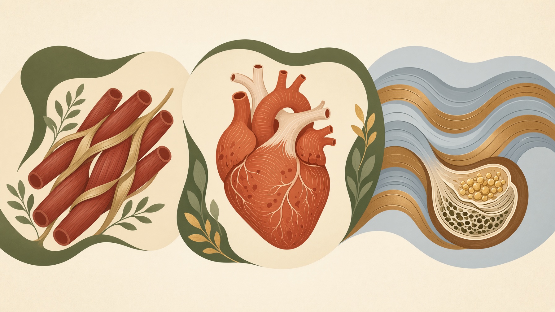
What Makes Skeletal, Cardiac, and Smooth Muscle Different?
Skeletal, cardiac, and smooth muscle share the ability to contract, but differences in cell structure and control suit each to different tasks.
Wikis
Skeletal-muscle fibers, also called myofibers, are the individual contractile cells that make up skeletal muscle. They are long, cylindrical cells that can extend for many centimeters and typically contain many nuclei positioned near the cell membrane. Each fiber is enclosed by a specialized plasma membrane called the sarcolemma and surrounded by a thin layer of connective tissue called the endomysium .1,2
Inside each muscle fiber are numerous myofibrils, long structures containing the proteins responsible for contraction. Myofibrils are organized into repeating units called sarcomeres, which contain thin filaments composed mainly of actin and thick filaments composed mainly of myosin. The regular arrangement of these filaments produces the characteristic striped, or striated, appearance of skeletal muscle .1,2
Muscle fibers are activated by motor neurons at specialized synapses called neuromuscular junctions. An electrical signal spreads along the sarcolemma and through inward extensions called T-tubules, triggering calcium release inside the fiber. The increased calcium allows actin and myosin to interact, generating force within the sarcomeres .2
An individual skeletal-muscle fiber should not be confused with an entire muscle. Fibers are grouped into bundles called fascicles, and many fascicles, together with connective tissues, nerves, and blood vessels, form a whole skeletal muscle. Fibers can also differ in their contractile and metabolic characteristics and are commonly classified into types such as Type I, Type IIa, and Type IIx .1,2
From this collection
Skeletal, cardiac, and smooth muscle share the ability to contract, but differences in cell structure and control suit each to different tasks.