
When people talk about “the abs,” they are usually referring to the most visible part of a much larger anatomical region. What appears from the outside as a single area is actually built from several muscles with different locations and relationships to one another. Looking beyond the familiar six-pack gives a much clearer picture of how the abdominal region is organized.
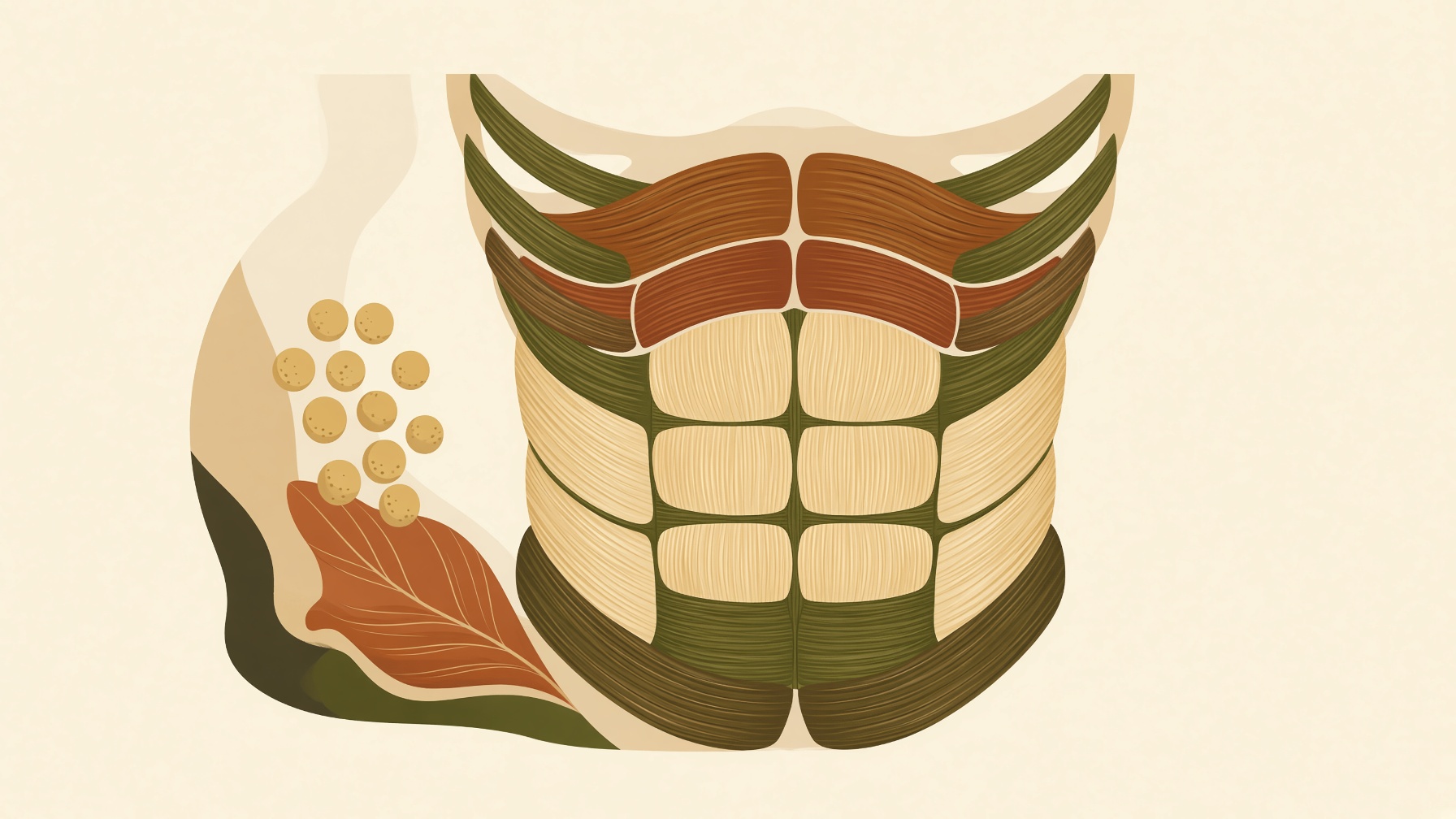
The major muscles discussed here are the external oblique, internal oblique, transversus abdominis, and rectus abdominis. Along the lateral abdominal wall, three of these form distinct layers. The external oblique is the most superficial, the internal oblique lies beneath it as the intermediate layer, and the transversus abdominis is the deepest of the three flat abdominal muscles .1
Those layers also differ in fiber orientation. External-oblique fibers generally run inferiorly and medially, while most internal-oblique fibers run superiorly and medially. Transversus-abdominis fibers are arranged predominantly across the abdomen. These different orientations contribute to multiple trunk movements, including rotation, although the three muscles do not contribute equally to rotation. Transversus abdominis is primarily important for compression and stabilization rather than functioning primarily as a rotational muscle. Together, the flat abdominal layers also help protect the internal abdominal organs .1
Rectus abdominis has a different arrangement. It sits medially in the anterior abdominal wall and is enclosed within the fibrous aponeurotic rectus sheath. The muscle is crossed by transverse fibrous bands called tendinous intersections. These divisions help create the familiar “six-pack” appearance when the muscle is visible. That appearance is not identical in everyone, however. Its visibility and exact pattern vary with individual anatomy, muscle development, and the amount of overlying adipose tissue .1,2
The anatomy relevant to trunk support also extends posteriorly. Muscles of the posterior abdominal wall, including structures such as quadratus lumborum and psoas major, contribute to positioning and movement of the trunk and lumbar region. That role adds another dimension to abdominal anatomy, showing that the structures involved in supporting the trunk are not confined to the muscles most visible from the front .1,3
Thinking beyond the idea of a single set of “abs” changes how the region makes sense anatomically. What is visible on the surface represents only part of a deeper arrangement of muscles and connective tissues that work across the trunk. That broader view is useful because the abdominal region is not designed solely for appearance; its anatomy reflects the many demands placed on the trunk during movement, stabilization, and everyday physical tasks.
Join the discussion
No account or email is required. Your display name and comment will be public.